Introduction: Why Diabetic Foot Ulcers Are a Serious Concern
Diabetes is a complex condition that affects millions of people worldwide, and one of its most serious complications is the development of foot ulcers. Diabetic foot ulcers are open sores or wounds that occur in approximately 15% of all people with diabetes, and they are among the leading causes of hospitalization and lower limb amputations globally. Despite being preventable in many cases, these ulcers can quickly escalate into life-threatening conditions if left untreated.
Understanding the causes, recognizing the symptoms early, and knowing the available treatment options can make the difference between a full recovery and a devastating outcome. Whether you are living with diabetes yourself or caring for someone who is, this comprehensive guide will walk you through everything you need to know about diabetic foot ulcers — from the biological mechanisms that cause them to the most advanced treatment strategies available today.
What Is a Diabetic Foot Ulcer?
A diabetic foot ulcer is a breakdown in the skin on the foot that exposes the tissues beneath it. These ulcers most commonly develop on the bottom of the foot, particularly under the big toe and the balls of the feet. They can range from shallow, surface-level wounds to deep ulcers that penetrate through layers of tissue, reaching muscle, tendons, and even bone.
What makes diabetic foot ulcers particularly dangerous is that people with diabetes often cannot feel pain in their feet due to nerve damage — a condition known as peripheral neuropathy. This means that an ulcer can develop and worsen significantly before the person even becomes aware that something is wrong. Combined with poor circulation and a compromised immune system, these ulcers can become infected rapidly and may result in gangrene or the need for amputation if not properly managed.
According to the American Diabetes Association, diabetic foot ulcers precede approximately 85% of diabetes-related amputations. This staggering statistic underscores why early detection, prevention, and prompt treatment are absolutely critical.
What Causes Diabetic Foot Ulcers?
Diabetic foot ulcers rarely have a single cause. Rather, they are the result of multiple interrelated factors that are directly or indirectly linked to the effects of prolonged high blood sugar levels on the body. Understanding these underlying causes is essential for both prevention and treatment.
1. Peripheral Neuropathy
One of the primary causes of diabetic foot ulcers is peripheral neuropathy, which is nerve damage that affects the feet and legs. High blood sugar levels over time can damage the nerve fibers throughout the body, but the longest nerves — those that reach the feet — are most vulnerable. This damage leads to a loss of sensation, making it impossible for a person to feel pain, heat, cold, or pressure in their feet.
As a result, minor injuries such as blisters, cuts, or pressure sores go unnoticed and untreated. Without the normal pain response that would prompt someone to seek care, these small wounds can quickly progress into full-blown ulcers.
2. Poor Circulation (Peripheral Artery Disease)
Diabetes significantly increases the risk of peripheral artery disease (PAD), a condition in which the arteries that supply blood to the legs and feet become narrowed or blocked due to the buildup of fatty deposits (atherosclerosis). Poor circulation means that the feet receive less oxygen and nutrients, both of which are essential for wound healing.
When a wound occurs in a person with compromised circulation, the body struggles to deliver the immune cells, growth factors, and nutrients necessary to heal the tissue. This dramatically slows the healing process and increases the risk of infection, allowing ulcers to persist and deepen over time.
3. High Blood Sugar Levels (Hyperglycemia)
Chronically elevated blood sugar levels — the hallmark of poorly controlled diabetes — play a central role in the development of foot ulcers. High glucose levels impair the function of white blood cells, which are the body’s primary defense against infection. This immunosuppressive effect means that even minor wounds are at a much greater risk of becoming seriously infected in a person with uncontrolled diabetes.
Additionally, hyperglycemia promotes inflammation and interferes with the normal cellular processes involved in wound repair, including collagen synthesis and cell migration. This creates an environment in which wounds simply cannot heal efficiently.
4. Foot Deformities and Abnormal Pressure Points
People with diabetes often develop structural changes in their feet over time. Neuropathy can affect the motor nerves that control the small muscles of the foot, leading to muscle weakness and imbalances that cause deformities such as hammertoes, claw toes, and Charcot foot — a condition characterized by the weakening of the bones in the foot, often leading to collapse of the arch.
These deformities create abnormal pressure points on the foot during walking and standing. Over time, this repeated pressure and friction causes the skin to break down, eventually forming an ulcer. Calluses often form at these pressure points and, if not properly managed, can themselves become sites of ulcer development.
5. Trauma and Minor Injuries
Because people with diabetic neuropathy cannot feel their feet properly, they are prone to accidental injuries that go unnoticed. Walking barefoot on a sharp object, wearing ill-fitting shoes that rub and create blisters, or even cutting toenails too short can result in wounds that, in a healthy person, would heal quickly and without complication. In a diabetic individual, however, these minor injuries can be the starting point of a serious ulcer.
6. Immune System Dysfunction
Diabetes impairs the overall function of the immune system in several ways. High blood sugar levels reduce the activity of neutrophils (a type of white blood cell critical for fighting bacterial infections), impair the function of macrophages that clear debris and coordinate wound healing, and reduce the effectiveness of cytokines that regulate the inflammatory response. This weakened immune response means that infections can take hold more easily and spread more rapidly in diabetic individuals.
7. Kidney Disease and Other Complications
Many people with long-standing diabetes also develop kidney disease (diabetic nephropathy), which further impairs the body’s ability to fight infections and heal wounds. The accumulation of waste products in the blood due to poor kidney function also contributes to immune dysfunction and poor tissue health, compounding the risk of foot ulcers.
Who Is Most at Risk?
While any person with diabetes can develop a foot ulcer, certain factors significantly increase the risk. Understanding these risk factors can help individuals and healthcare providers take targeted preventive measures.
- Type 1 or Type 2 diabetes with poor blood sugar control — The longer blood sugar remains elevated, the greater the accumulated damage to nerves and blood vessels.
- Diabetes duration — People who have had diabetes for 10 years or more have a significantly higher risk due to cumulative nerve and vascular damage.
- History of previous foot ulcers or amputations — Prior ulcers are one of the strongest predictors of future ulcers.
- Peripheral neuropathy with sensory loss — Loss of protective sensation dramatically increases vulnerability to injury.
- Peripheral artery disease — Compromised blood flow is a major risk factor for both ulcer development and poor healing.
- Smoking — Nicotine constricts blood vessels and worsens circulation, compounding the effects of PAD.
- Obesity — Excess body weight increases pressure on the feet and exacerbates insulin resistance.
- Poor foot hygiene or footwear habits — Going barefoot, wearing ill-fitting shoes, or neglecting foot care increases exposure to injury.
- Visual impairment — People who cannot see well may be unable to inspect their feet for signs of injury or infection.
- Living alone or having limited access to healthcare — Social isolation and lack of medical care can delay diagnosis and treatment.
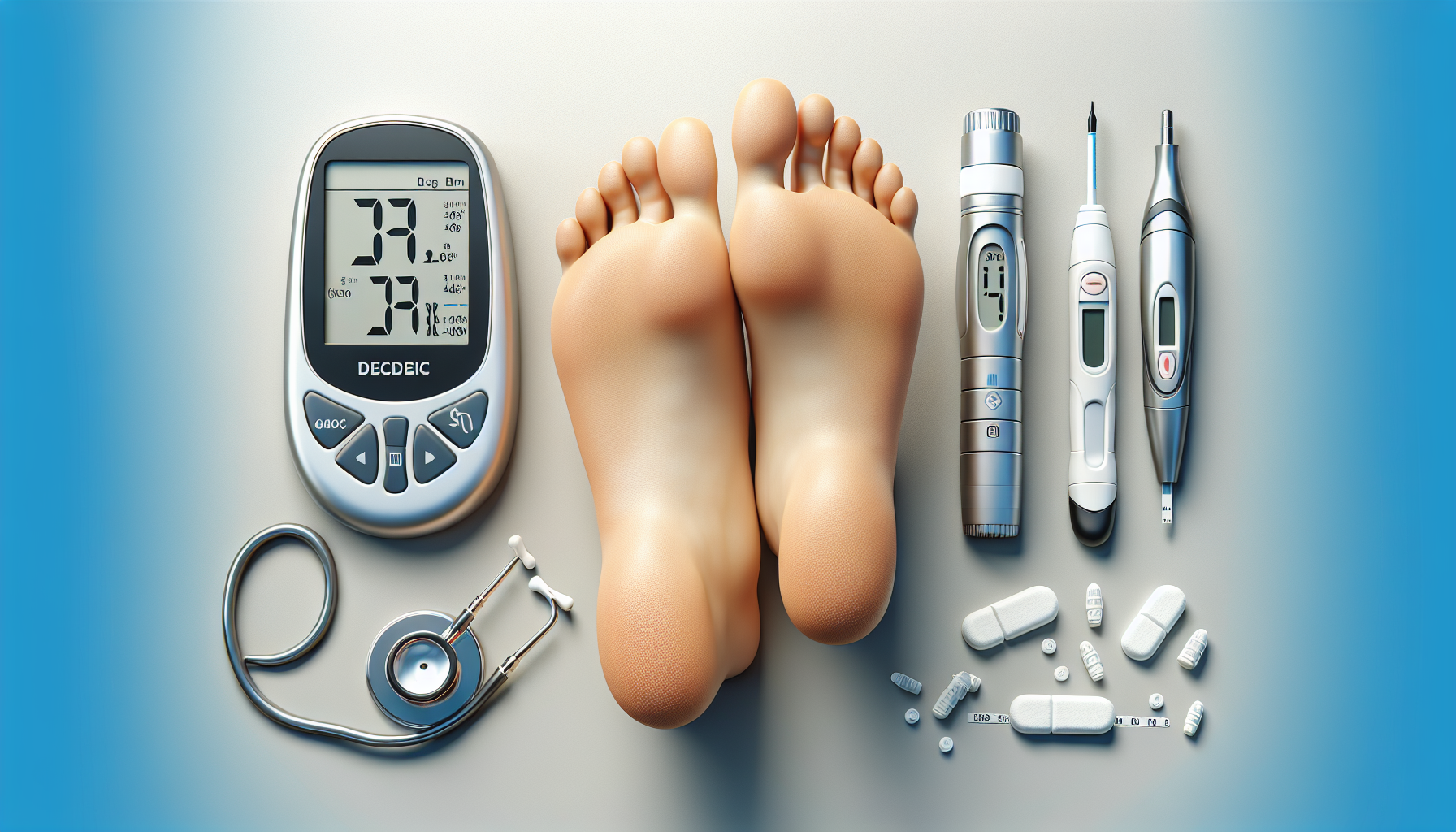
Symptoms of Diabetic Foot Ulcers
Because neuropathy can mask pain, recognizing the visual and other sensory symptoms of a diabetic foot ulcer is critically important. Regular foot inspections — both self-examinations and professional assessments — are key to early detection.
Early Warning Signs
- Redness, swelling, or warmth in a specific area of the foot
- Skin discoloration — areas of the skin turning white, brown, or black
- Calluses or corns with dried blood inside them
- Blisters, cuts, or cracks in the skin that are not healing as expected
- Drainage — unusual fluid, pus, or blood staining socks or shoes
- Unpleasant odor coming from the foot
- Thickening or hardening of the skin in pressure-prone areas
Symptoms of an Established Ulcer
- An open sore or wound on the foot, particularly on the sole or around the toes
- Visible tissue, tendons, or bone at the base of the wound in severe cases
- Black or necrotic tissue surrounding the wound (eschar), indicating gangrene
- Fever, chills, or general malaise — signs of systemic infection
- Swelling of the entire foot or leg
- Increased blood sugar levels that are difficult to control, often indicating underlying infection
When to Seek Immediate Medical Attention
If you notice any of the following, seek emergency medical care immediately:
- An ulcer that has turned black or has a foul-smelling discharge
- Rapidly spreading redness or red streaks extending up the leg (signs of cellulitis or septicemia)
- High fever or chills accompanied by a foot wound
- Loss of consciousness or confusion in someone with diabetes and a foot wound
These are signs of serious, potentially life-threatening infection that requires immediate hospitalization and treatment.
Classification of Diabetic Foot Ulcers
Healthcare providers use classification systems to assess the severity of diabetic foot ulcers and guide treatment decisions. The most widely used system is the Wagner Grading Scale, which classifies ulcers on a scale from 0 to 5:
- Grade 0: No open lesions; skin is intact but foot may have deformities or areas of pre-ulcerative changes such as calluses.
- Grade 1: Superficial ulcer involving only the skin surface, without infection or deep tissue involvement.
- Grade 2: Deeper ulcer extending to ligament, tendon, joint capsule, or deep fascia, without bone involvement or abscess.
- Grade 3: Deep ulcer with abscess, osteomyelitis (bone infection), or joint infection.
- Grade 4: Gangrene (tissue death) limited to the toes or forefoot.
- Grade 5: Extensive gangrene involving the entire foot, requiring amputation.
Another commonly used system is the University of Texas Wound Classification System, which adds variables for the presence of infection and ischemia (insufficient blood supply) to give a more nuanced picture of ulcer severity.
Diagnosis of Diabetic Foot Ulcers
Diagnosis of a diabetic foot ulcer involves a thorough clinical assessment combined with various diagnostic tests to evaluate the extent of the wound, the presence of infection, blood flow, and nerve function.
Physical Examination
A healthcare provider will visually examine the ulcer, assessing its size, depth, location, and the condition of surrounding tissue. They will check for signs of infection such as redness, warmth, swelling, and discharge. The examination also includes assessment of the overall foot structure for deformities that may be contributing to the ulcer.
Neurological Assessment
Tests such as the Semmes-Weinstein monofilament test are used to assess protective sensation in the foot. A thin nylon filament is pressed against different areas of the foot, and if the patient cannot feel it, significant neuropathy is present.
Vascular Assessment
The ankle-brachial index (ABI) is a simple non-invasive test that compares blood pressure in the ankle with blood pressure in the arm to detect peripheral artery disease. Doppler ultrasound or arteriography may also be performed to evaluate blood flow more precisely.
Laboratory Tests
Blood tests are used to assess overall health, blood sugar control (HbA1c levels), kidney function, and inflammatory markers such as C-reactive protein (CRP) and white blood cell count. Wound swabs or tissue cultures are taken to identify the specific bacteria causing an infection and determine which antibiotics will be most effective.
Imaging Studies
X-rays are used to detect gas in the tissues (a sign of severe infection), foreign bodies, or bone changes. MRI (Magnetic Resonance Imaging) is the most sensitive imaging tool for detecting osteomyelitis (bone infection), which is a serious complication that significantly changes the treatment approach.
Treatment of Diabetic Foot Ulcers
Treatment for diabetic foot ulcers is multifaceted and typically requires a team of healthcare professionals, including endocrinologists, podiatrists, vascular surgeons, infectious disease specialists, and wound care nurses. The goals of treatment are to promote healing, prevent or treat infection, restore blood flow, and prevent recurrence.
1. Blood Sugar Control
Effective management of blood glucose levels is the cornerstone of diabetic foot ulcer treatment. Without good blood sugar control, even the most advanced wound care interventions will be less effective. Target HbA1c levels, medication adjustments, dietary changes, and regular monitoring are all essential components of this aspect of care.
2. Wound Debridement
Debridement is the removal of dead, infected, or foreign tissue from a wound to promote the growth of healthy new tissue. There are several methods of debridement used in the management of diabetic foot ulcers:
- Sharp/Surgical debridement: A surgeon or wound care specialist uses a scalpel or scissors to cut away dead tissue. This is the most rapid and thorough method.
- Enzymatic debridement: Topical agents containing enzymes are applied to the wound to chemically break down dead tissue.
- Autolytic debridement: Moisture-retentive dressings are used to allow the body’s own enzymes to dissolve dead tissue naturally.
- Mechanical debridement: Techniques such as wet-to-dry dressings or wound irrigation are used to physically remove dead tissue.
- Maggot therapy: Medical-grade maggots are applied to the wound to selectively consume dead tissue while leaving healthy tissue intact — a surprisingly effective and evidence-based treatment.
3. Wound Dressings
Choosing the right wound dressing is critical for creating the optimal environment for healing. The type of dressing used depends on the characteristics of the wound, including its depth, the amount of drainage, and the presence of infection. Common dressing types include