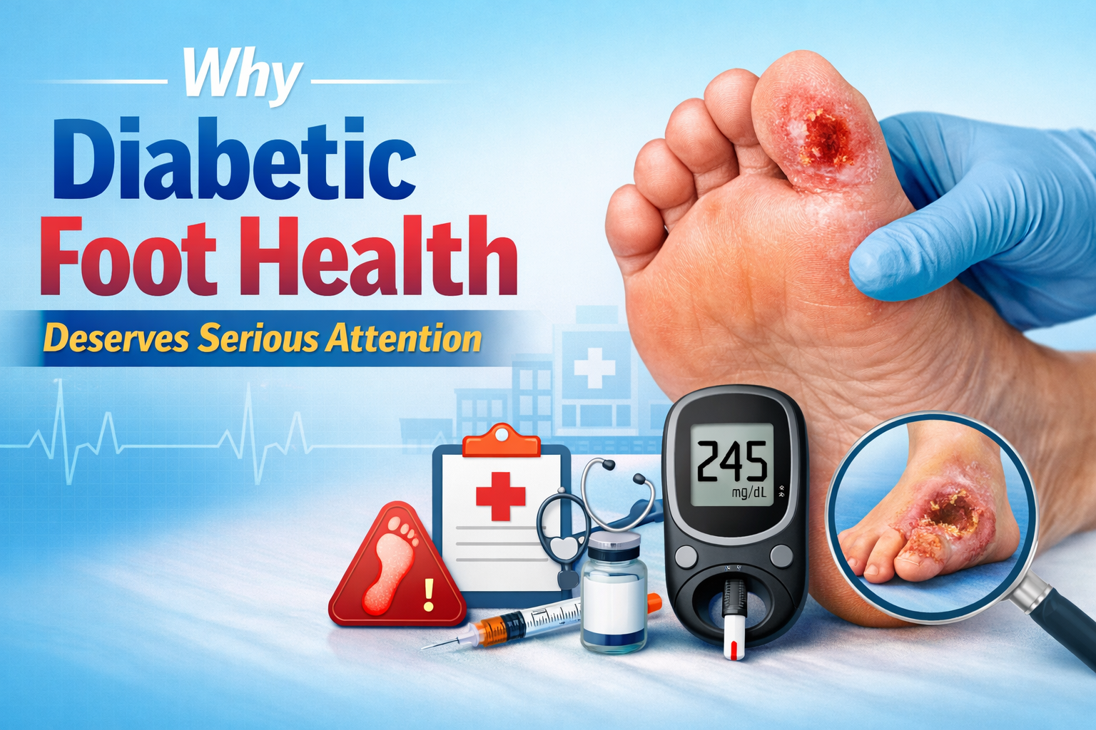
Introduction: Why Diabetic Foot Ulcers Demand Serious Attention
Diabetic foot ulcers are among the most serious and potentially life-altering complications that people living with diabetes can face. These open wounds, which most commonly develop on the bottom of the foot, affect approximately 15% of all people with diabetes at some point during their lifetime. What may begin as a small blister, callus, or minor cut can rapidly escalate into a deep, infected wound that is stubbornly resistant to healing.
The stakes could not be higher. Diabetic foot ulcers are the leading cause of non-traumatic lower limb amputations worldwide, accounting for more than 80% of all diabetes-related amputations. Beyond the physical toll, these wounds place an enormous emotional, financial, and social burden on patients and their families. Hospital stays are prolonged, quality of life diminishes, and the psychological weight of a chronic, non-healing wound can be devastating.
Yet, with the right knowledge, a proactive approach, and a comprehensive wound care strategy, many diabetic foot ulcers can be successfully treated — and even prevented altogether. This guide is designed to walk you through everything you need to know about wound care for diabetic foot ulcers, from understanding why they occur to the most advanced treatment options available today.
Understanding Diabetic Foot Ulcers: The Basics
What Is a Diabetic Foot Ulcer?
A diabetic foot ulcer (DFU) is an open sore or wound that occurs in approximately 15% of patients with diabetes and is commonly located on the bottom of the foot. These ulcers develop as a result of a combination of factors unique to the diabetic condition, including nerve damage, poor circulation, and a compromised immune system. Unlike a typical wound in a healthy individual, a diabetic foot ulcer often heals slowly — or not at all — without proper intervention.
Why Do Diabetic Foot Ulcers Develop?
To understand wound care for diabetic foot ulcers, it’s essential to first understand the underlying mechanisms that cause them. Several key factors contribute to their development:
- Peripheral Neuropathy: High blood sugar levels over time damage the nerves in the feet and legs. This nerve damage, known as peripheral neuropathy, causes loss of sensation — meaning patients cannot feel pain, pressure, or temperature changes in their feet. A small pebble in a shoe, a blister from ill-fitting footwear, or a minor cut can go completely unnoticed until it has already become a serious wound.
- Peripheral Arterial Disease (PAD): Diabetes damages blood vessels, reducing blood flow to the extremities. Poor circulation means that oxygen and nutrients cannot reach the tissues in the feet effectively, dramatically impairing the body’s natural ability to heal wounds.
- Immune System Dysfunction: Elevated blood glucose levels impair the function of white blood cells, making it harder for the body to fight off infection. This increases the risk that even minor wounds will become infected.
- Foot Deformities: Conditions such as hammertoes, bunions, and Charcot foot (a complication of neuropathy that causes bone and joint damage) can create abnormal pressure points on the foot, leading to skin breakdown and ulceration.
- Dry Skin and Calluses: Neuropathy also affects the sweat glands in the feet, leading to excessively dry skin that cracks easily. Calluses that build up over pressure points can also break down and form ulcers.
How Are Diabetic Foot Ulcers Classified?
Healthcare providers use classification systems to assess the severity of diabetic foot ulcers and guide treatment decisions. The most widely used system is the Wagner Grading System, which classifies ulcers on a scale of 0 to 5:
- Grade 0: No open lesion; high-risk foot with intact skin
- Grade 1: Superficial ulcer involving the full thickness of the skin but not underlying tissues
- Grade 2: Deep ulcer penetrating to tendon, capsule, or bone
- Grade 3: Deep ulcer with abscess, osteomyelitis, or joint sepsis
- Grade 4: Gangrene of the forefoot
- Grade 5: Gangrene of the entire foot
Another commonly used tool is the University of Texas Wound Classification System, which assesses depth, infection, and ischemia (reduced blood flow). Both systems help clinicians determine the most appropriate course of treatment.
Step-by-Step Wound Care for Diabetic Foot Ulcers
Proper wound care is the cornerstone of diabetic foot ulcer management. The goals of wound care are to promote healing, prevent infection, and protect the surrounding healthy tissue. Here is a comprehensive overview of the essential steps involved:
Step 1: Assessment and Diagnosis
Before any treatment begins, a thorough assessment of the wound and the patient’s overall health is essential. This includes:
- Measuring the wound’s size, depth, and location
- Assessing the wound bed for signs of infection, necrosis (dead tissue), or healthy granulation tissue
- Evaluating circulation in the affected limb using tools like ankle-brachial index (ABI) measurements or Doppler ultrasound
- Checking for signs of infection such as redness, warmth, swelling, pain, or discharge
- Reviewing the patient’s blood glucose levels and overall diabetes management
A proper assessment sets the foundation for an individualized treatment plan. Self-assessment at home is also critically important — patients should inspect their feet daily and report any changes to their healthcare provider immediately.
Step 2: Debridement — Removing Dead and Damaged Tissue
One of the most critical components of diabetic foot ulcer wound care is debridement, the process of removing dead, damaged, or infected tissue from the wound. Dead tissue (necrotic tissue or slough) creates an environment that harbors bacteria and prevents healthy tissue from forming. There are several methods of debridement:
- Sharp or Surgical Debridement: A healthcare professional uses a scalpel, scissors, or curette to manually remove devitalized tissue. This is the fastest and most effective method and is often the first choice for clinicians.
- Autolytic Debridement: This method uses the body’s own enzymes and moisture to soften and break down dead tissue. It is achieved through the use of moisture-retentive dressings such as hydrogels or hydrocolloids. It is gentle but slower than surgical methods.
- Enzymatic Debridement: Topical agents containing enzymes (such as collagenase) are applied to the wound to chemically break down necrotic tissue. This approach is useful when sharp debridement is not feasible.
- Mechanical Debridement: Techniques such as wet-to-dry dressings, wound irrigation, or hydrotherapy remove dead tissue mechanically. However, these methods can also damage healthy tissue and are less commonly used today.
- Biological Debridement (Maggot Therapy): Medical-grade maggots are applied to the wound, where they consume necrotic tissue and bacteria. While it may sound unusual, this method is highly effective for wounds that have not responded to other treatments.
Regular, repeated debridement is often necessary as the wound heals. Studies show that wounds treated with frequent debridement heal significantly faster than those treated less frequently.
Step 3: Infection Control and Management
Infection is one of the most dangerous complications of diabetic foot ulcers and must be addressed aggressively. Because the diabetic immune system is compromised, even minor wound contamination can escalate into a serious systemic infection.
Signs of infection in a diabetic foot ulcer include:
- Increased redness, warmth, or swelling around the wound
- Purulent (pus-like) discharge
- Foul odor
- Increased pain or tenderness (even in patients with neuropathy, some sensation may be retained)
- Fever, chills, or elevated white blood cell count
- Rapid increase in wound size
Treatment for infected diabetic foot ulcers typically includes:
- Wound Cultures: Before initiating antibiotic therapy, wound cultures are taken to identify the specific bacteria causing the infection, allowing for targeted antibiotic selection.
- Topical Antiseptics: Agents such as cadexomer iodine, silver-containing dressings, or medical-grade honey may be used to reduce bacterial burden in the wound. These are particularly useful for managing biofilms — communities of bacteria that are resistant to conventional antibiotics.
- Systemic Antibiotics: For moderate to severe infections, oral or intravenous antibiotics are prescribed based on culture results. Common organisms include Staphylococcus aureus (including MRSA), Streptococcus species, and gram-negative bacteria.
- Surgical Intervention: In cases of deep infection, abscess formation, or osteomyelitis (bone infection), surgical drainage, removal of infected bone, or amputation of affected toes may be necessary.
Step 4: Choosing the Right Wound Dressing
Selecting the appropriate wound dressing is a critical and nuanced part of diabetic foot ulcer care. The ideal dressing should maintain a moist wound environment (which promotes healing), absorb excess exudate, protect against contamination, and support the healing process without causing further trauma upon removal.
There is no single “best” dressing for all diabetic foot ulcers — the choice depends on the characteristics of the wound, including its depth, the amount of exudate, and the presence of infection. Here is an overview of common dressing types:
- Hydrogel Dressings: These moisture-donating dressings are ideal for dry wounds and support autolytic debridement. They help rehydrate necrotic tissue and provide a cool, soothing environment.
- Hydrocolloid Dressings: Suitable for wounds with minimal to moderate exudate, these dressings create a moist, occlusive environment and can stay in place for several days, reducing the frequency of dressing changes.
- Foam Dressings: Highly absorbent and available in a variety of forms, foam dressings are ideal for wounds with moderate to heavy exudate. They can be used with or without adhesive borders.
- Alginate Dressings: Derived from seaweed, alginate dressings are extremely absorbent and are used for heavily exuding wounds. They form a gel upon contact with wound fluid, maintaining moisture while absorbing excess drainage.
- Silver-Impregnated Dressings: These dressings release silver ions that have broad-spectrum antimicrobial properties. They are used for infected or at-risk wounds to reduce bacterial burden.
- Collagen Dressings: Collagen dressings promote wound healing by attracting new cells into the wound bed and stimulating the formation of new tissue. They are often used for chronic, slow-healing wounds.
- Antimicrobial Dressings with Cadexomer Iodine or PHMB: Polyhexamethylene biguanide (PHMB) and cadexomer iodine dressings are effective against a broad spectrum of bacteria, including biofilm-forming organisms, and are commonly used in chronically infected ulcers.
Dressings should be changed according to the manufacturer’s instructions and the clinical condition of the wound. Over-frequent changes can disturb the healing process, while infrequent changes can lead to maceration and infection.
Step 5: Offloading — Taking Pressure Off the Wound
Offloading is arguably the single most important factor in healing diabetic foot ulcers located on the plantar (bottom) surface of the foot. Continued pressure on an ulcer prevents healing and can cause the wound to worsen rapidly, no matter how excellent the wound care applied.
The gold standard for offloading plantar diabetic foot ulcers is the Total Contact Cast (TCC). A TCC is a specially applied cast that distributes pressure evenly across the entire plantar surface of the foot, dramatically reducing pressure on the ulcer site. Research consistently shows that TCC achieves healing rates of 72–100% for neuropathic plantar ulcers.
Other offloading methods include:
- Removable Cast Walkers (RCWs): Also known as CAM boots or walking boots, these devices provide effective offloading but are less effective than TCCs because patients often remove them, reducing compliance.
- Irremovable Cast Walkers: When a RCW is made irremovable (by wrapping it with a layer of fiberglass), it achieves offloading outcomes comparable to TCC.
- Half Shoes and Forefoot Relief Shoes: These specialized shoes unload pressure from the front of the foot and are often used for forefoot ulcers.
- Custom Orthotic Insoles: Pressure-redistributing insoles can help manage plantar pressure and are often used in the prevention of recurrence once the ulcer has healed.
- Bed Rest and Wheelchair Use: In severe cases, complete rest from weight-bearing may be necessary, particularly when infection or ischemia is present.
Step 6: Blood Glucose Management
Optimal blood glucose control is fundamental to wound healing in diabetic foot ulcers. Elevated blood sugar impairs virtually every aspect of the healing process, including immune function, collagen production, and blood vessel formation (angiogenesis). Research shows that patients with poorly controlled diabetes have significantly worse ulcer healing outcomes compared to those with well-managed blood sugar levels.
Goals for blood glucose management in the context of wound healing typically include:
- Maintaining HbA1c levels below 7–8% (as advised by the treating physician)
- Keeping fasting blood glucose levels within the target range set by the healthcare team
- Working closely with an endocrinologist or diabetes care team to adjust medications as needed
- Adopting a diabetes-friendly diet rich in nutrients that support wound healing, including protein, vitamin C, zinc, and vitamin D
Advanced Therapies for Diabetic Foot Ulcers
When standard wound care measures are insufficient, several advanced therapies have been developed to stimulate healing in stubborn or complex diabetic foot ulcers. These treatments are typically used in conjunction with — not as a replacement for — the fundamental steps outlined above.
Negative Pressure Wound Therapy (NPWT)
Also known as vacuum-assisted closure (VAC therapy), NPWT involves applying a sealed wound dressing connected to a vacuum pump that applies continuous or intermittent negative pressure to the wound surface. This therapy promotes healing by:
- Removing excess fluid and infectious material from the wound
- Reducing wound edema
- Stimulating blood flow to the wound area
- Encouraging granulation tissue formation
- Drawing wound edges together